
Advanced Dental Technology
Modern dentistry is not simply about better equipment. It is about using the right technology at the right stage of care, to see more clearly, plan more precisely, treat more conservatively and make your experience more comfortable.
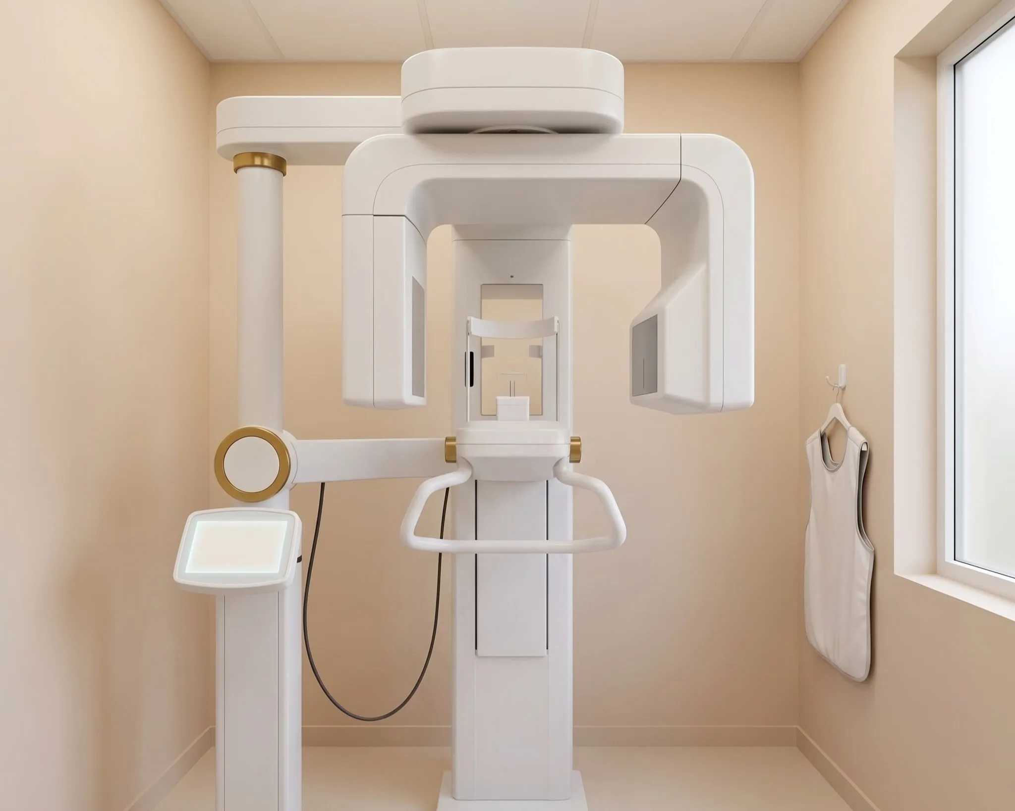
CBCT 3D Imaging
Cone Beam Computed Tomography (CBCT) creates detailed three-dimensional images of the teeth, roots, jawbone and surrounding anatomical structures. This provides a more comprehensive view than conventional two-dimensional dental imaging in situations where 3D information is clinically appropriate.
CBCT imaging can support treatment planning for dental implants, complex root canal cases, oral and maxillofacial surgery, impacted teeth and evaluation of available bone and anatomical structures.
By allowing the clinical team to assess anatomy from multiple angles, 3D imaging can contribute to more informed diagnosis and treatment planning.
Digital Intraoral Scanning
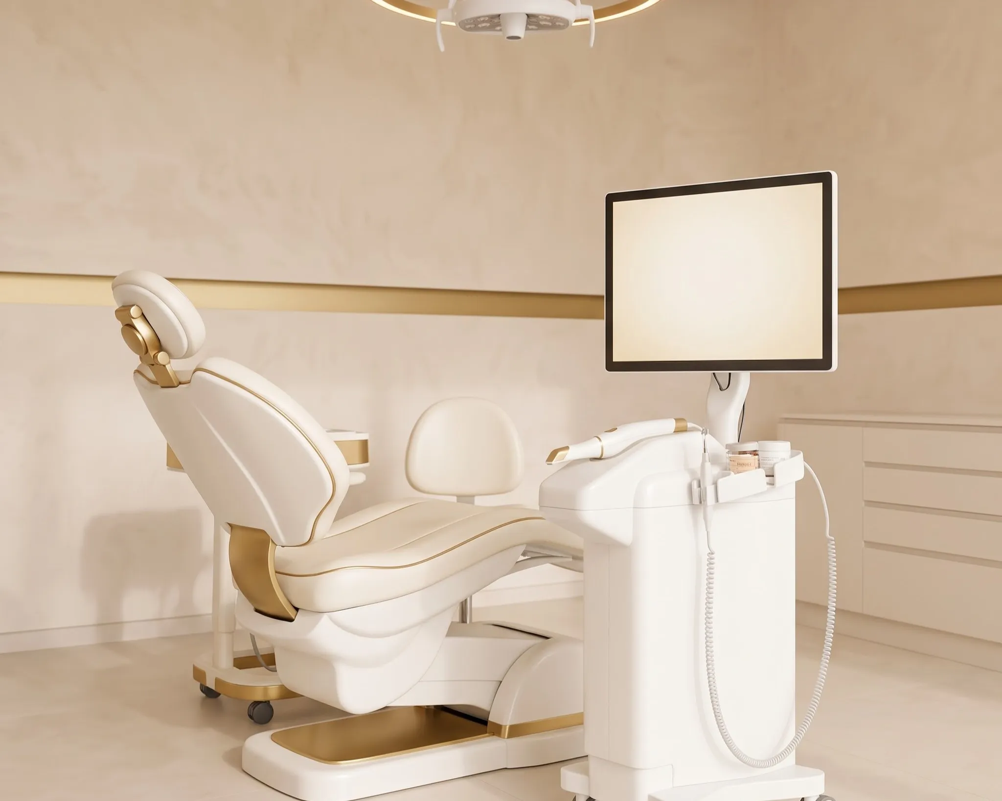
Digital intraoral scanners capture highly detailed three-dimensional images of the teeth and surrounding oral structures, creating a digital record without the need for traditional impression materials in appropriate cases.
These digital scans can support a variety of modern dental workflows, including crowns, bridges, aligners, implant planning, restorative dentistry and smile design.
The digital format also makes it easier for the dental team to review the patient's teeth, explain findings and communicate treatment information where required.
Aurea's existing treatment approach already incorporates intraoral scanning as part of digital smile assessment and planning.
Dental Operating Microscope
A dental operating microscope provides high magnification and enhanced illumination, allowing clinicians to visualise extremely fine dental structures that may be difficult to see with the naked eye.
Microscopic dentistry is particularly valuable for complex root canal treatments, endodontic retreatment, locating additional canals, identifying fine anatomical details and managing challenging dental procedures.
Greater magnification can help the clinician work with improved visual control when precision is especially important.
A microscope-assisted approach is particularly aligned with Aurea's endodontic focus and precision-driven philosophy.
Digital Smile Design
Digital Smile Design brings together digital photographs, intraoral scans and treatment-planning information to evaluate the appearance and proportions of a smile before appropriate cosmetic treatment is carried out.
It can support the planning of treatments such as smile makeovers, veneers, crowns and aesthetic restorations, helping the clinical team evaluate tooth proportions, symmetry and the relationship between the smile and the surrounding facial features.
Digital visualisation can also make treatment discussions more collaborative by helping patients better understand the proposed aesthetic direction.
Digital Dental X-Rays
Digital dental X-rays provide detailed images of areas that cannot always be evaluated adequately through a visual examination alone.
They can assist clinicians in assessing teeth, roots, bone levels, cavities, infections and other underlying dental conditions, depending on the type of image required.
Digital imaging also allows diagnostic information to be reviewed efficiently during the consultation and incorporated into the overall clinical assessment.
Computer-Guided Implant Planning
Digital implant planning combines diagnostic imaging, 3D information and virtual planning tools to evaluate the proposed position of a dental implant in relation to the available bone and surrounding anatomical structures.
For appropriate cases, computer-assisted planning can support a more precise evaluation of implant position, angulation, depth and restorative considerations before the surgical procedure.
This planning-focused approach is particularly valuable when replacing missing teeth requires careful coordination between surgical and restorative considerations.
Intraoral Photography
Intraoral photography provides high-resolution images of specific areas inside the mouth, allowing dental conditions and treatment requirements to be documented and reviewed more clearly.
Photographs can help patients understand issues such as tooth wear, fractures, discolouration, gum conditions, restorative concerns and other visible dental findings.
They can also support treatment documentation and communication throughout the patient's dental journey.
Digital Endodontic Technology
Contemporary endodontic treatment combines digital diagnostics with specialised instruments and technology designed to improve control and accuracy throughout root canal procedures.
Depending on the clinical situation, digital endodontic workflows may incorporate advanced imaging, electronic working-length measurement, rotary or reciprocating instrumentation and magnification.
These technologies work together to support careful cleaning, shaping and treatment of the root canal system while helping clinicians manage complex endodontic cases more effectively.